
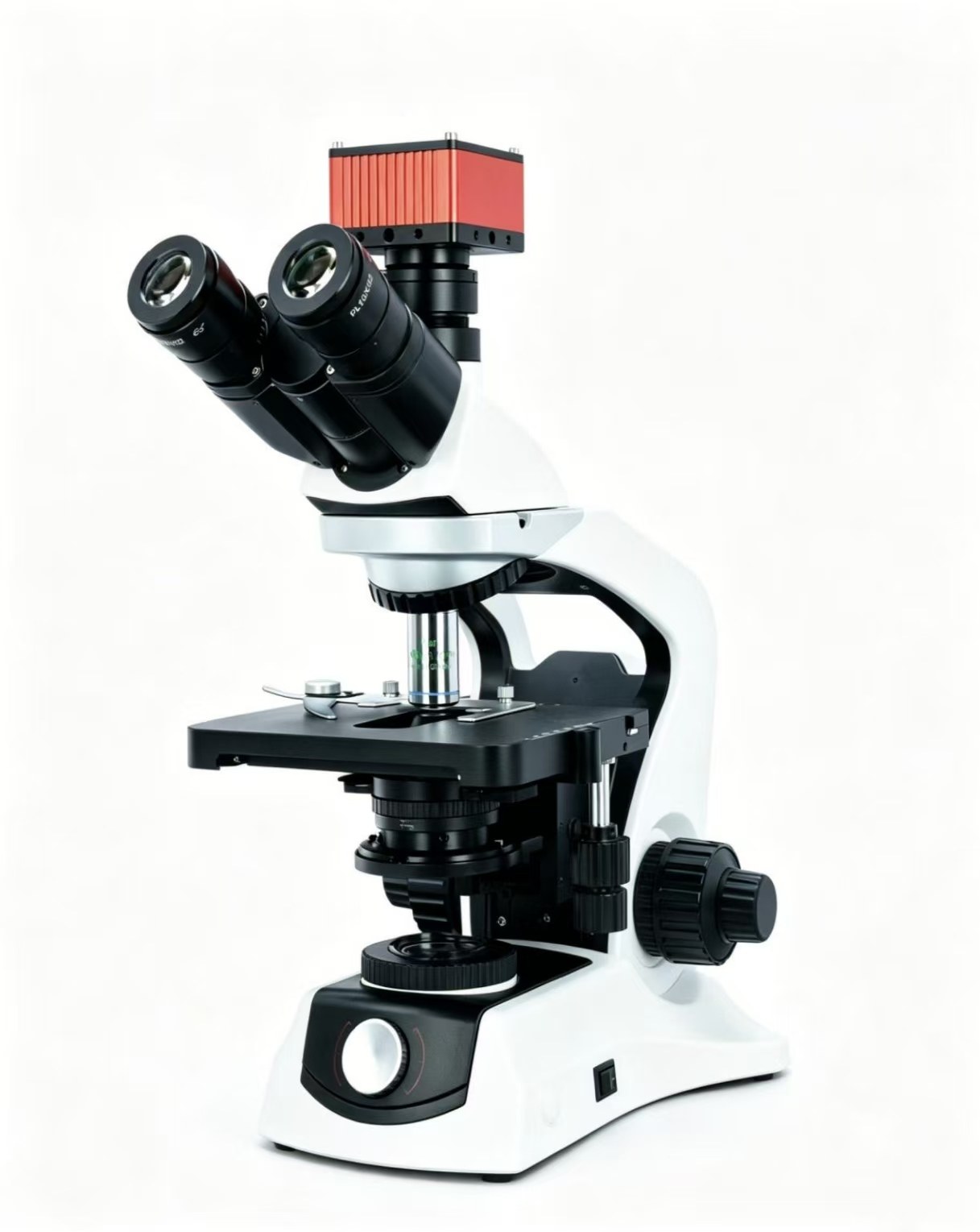
YS600
Darkfield
- 40× plan achromatic objective
- Darkfield condenser
- Dedicated darkfield configuration
- Compatible with HD3900
Microscopy
Biological microscope series
Choose darkfield, phase contrast, or switchable darkfield and phase contrast. All three can be paired with the HD3900 camera for on-screen viewing, capture and documentation.
Darkfield
Phase contrast
Dual-mode flagship
| Eyepieces | High-eyepoint, widefield plan eyepieces |
|---|---|
| Viewing head | 30° inclined trinocular head |
| Focusing | Low-position coaxial coarse / fine focusing; 0.005 mm fine-focus precision |
| Mechanical stage | 210 × 140 mm; X/Y travel 75 × 50 mm |
| Illumination | 5 W LED transmitted illumination |
| Interpupillary distance | 48–75 mm |
| Diopter adjustment | ±5 |
| Camera interface | C-mount |
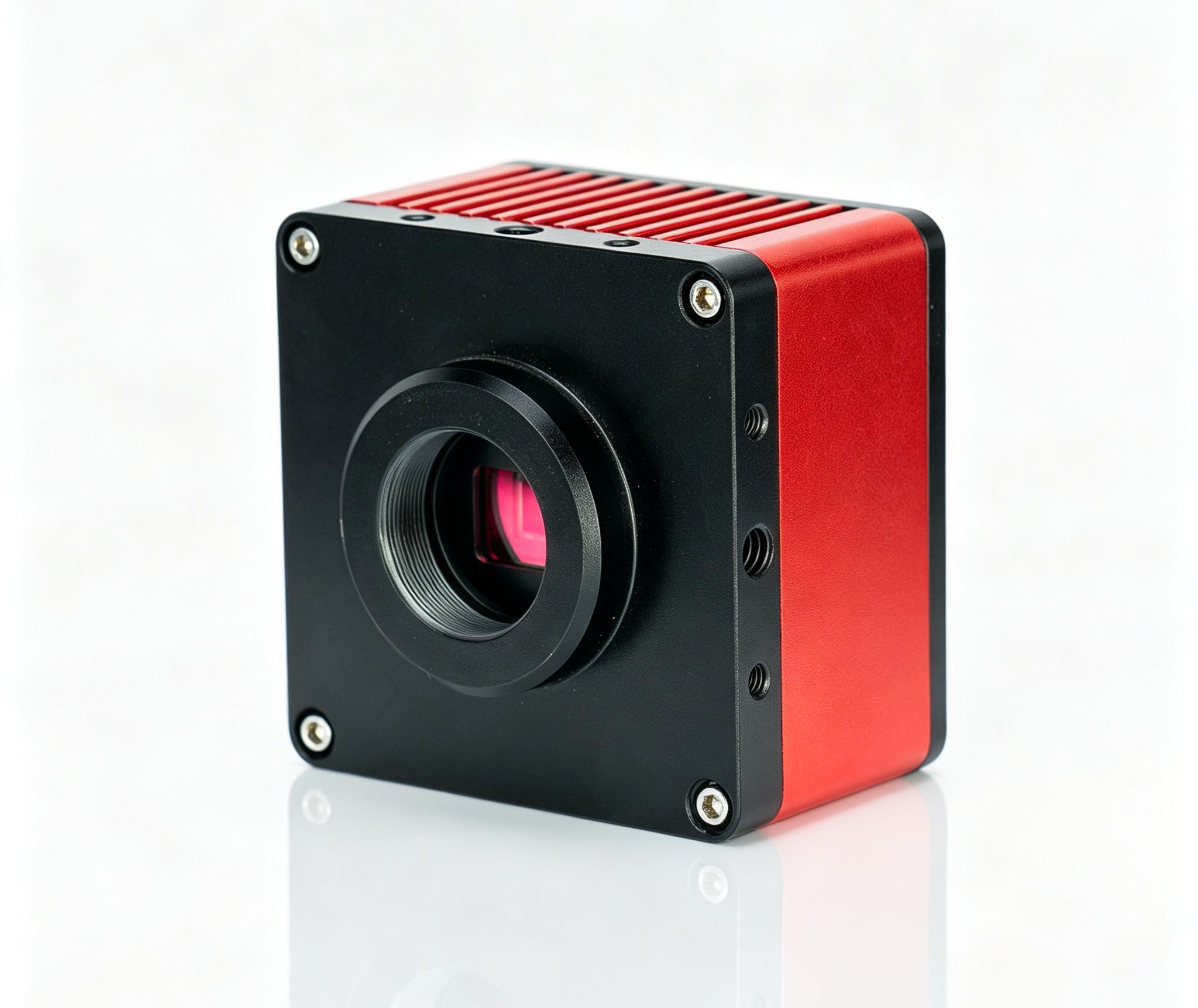
1080p standard · 4K optional · 60 fps
Selection guide
How microscopes are used in clinical testing, life science, food inspection, materials analysis, semiconductors and industrial inspection, and how to choose magnification, numerical aperture, working distance and illumination.
Microscopes are used across medicine, research, industry and teaching. The requirements change with the task. Cell observation depends on resolution and contrast. Circuit-board rework depends on working distance and a stereo view. Materials measurement depends on calibration, accuracy and repeatability.
The figures below are common configurations and a starting point for selection. They are not a uniform minimum requirement, and they are not a performance claim for a particular product. The final configuration should follow the sample, the observation goal, the method and any applicable standard.
Swipe sideways to see the full table.
| Application | Microscope type often used | Typical total magnification | Parameters and configuration notes |
|---|---|---|---|
| Histopathology sections | Upright brightfield microscope | 40–400× | Often 4×, 10×, 20× and 40× objectives. Flat-field imaging, colour rendering, even illumination and stage handling matter. |
| Stained blood films and bacterial smears | Upright biological microscope | 400–1000× | Often 40× and a 100× oil-immersion objective. A 100× oil objective commonly has an NA of about 1.25 or higher. Detail and faithful stain colour matter. |
| Routine samples such as urine sediment | Brightfield or phase-contrast microscope | 100–400× | Often 10× and 40×. Phase contrast helps transparent, low-contrast samples. Field of view and focus stability matter. |
| Cell-culture observation | Inverted phase-contrast microscope | 100–400× | Often 10×, 20× and 40×. Working distance must suit the vessel. Check the vessel-bottom thickness and objective compatibility. |
| Fluorescence localisation and 3D tissue imaging | Fluorescence or confocal microscope | 100–1000× | Match excitation, filters and detection channels to the dye. Fine imaging often uses objectives with an NA of about 0.75–1.4. Weak signals need detection sensitivity. 3D imaging needs optical sectioning and Z control. |
| Long-term live-cell observation | Inverted imaging system with environmental control | 100–600× | Temperature, gas, humidity, autofocus and low phototoxicity matter. Mammalian cells are often held near 37 °C. CO₂ depends on the culture system. Acquisition speed follows the experiment. |
| Bacteria, yeast and mould | Biological microscope, with a stereo microscope when needed | Yeast and mould often 100–400×; bacterial morphology often 1000× | Bacterial morphology often needs a 100× oil objective. Phase contrast suits transparent living samples. A stereo microscope can show whole colonies. Species identification usually also needs other methods. |
| Food foreign bodies, fibres and particles | Stereo, polarising or biological microscope | Stereo about 10–80×; fine particles about 100–400× | Reflected and transmitted light, working distance, field of view and size calibration matter. Polarisation helps fibre and crystal work. |
| Grains, inclusions and cracks | Metallurgical microscope | 50–1000× | Opaque samples use reflected light. Objectives are often 5× to 100×. Choose brightfield, darkfield, polarisation or DIC for the task. Quantitative work needs controlled preparation, calibration and analysis software. |
| Wafer surfaces and micrometre-scale defects | Reflected-light industrial microscope | 50–1000× | High-NA objectives, brightfield and darkfield, DIC, stage travel and positioning repeatability matter. Finer structure may need an electron microscope. |
| PCB and solder inspection and rework | Stereo or digital microscope | Routine inspection 5–30×; detail up to about 80× | Long working distance, a wide field, a stereo view and adjustable lighting come first. Rework can start from a working distance of about 100 mm or more. Video work needs low latency. |
| Precision parts, burrs and wear | Stereo, metallurgical or 3D measuring microscope | Routine about 10–200×; smaller defects may need higher magnification | Match working distance, depth of field, lighting and stage space to the sample. Measurement needs a stated calibration method, accuracy and repeatability. |
| Coatings, step height and surface topography | 3D measuring microscope | Set by the measuring range and the objective | Compare lateral and Z measuring ability, range, repeatability, surface compatibility and the method. Do not select on magnification alone. |
| Algae, plankton and particles in water | Upright or inverted biological microscope | 100–400×; detail up to 1000× | Phase contrast helps low-contrast samples. Counting needs a suitable chamber and calibration. Sedimentation counting needs an inverted layout and vessel compatibility. |
| Mineral and rock thin sections | Polarising microscope | 25–400× | Polariser, analyser and a rotating stage. Add compensators, a Bertrand lens and conoscopic accessories for the identification. |
| Hair, fibres, pigments and trace marks | Stereo, comparison, polarising or fluorescence microscope | Overview about 10–80×; fine structure about 100–400× | Consistent lighting, colour recording, polarisation and image calibration matter. Side-by-side comparison can use a comparison microscope. |
| Teaching and outreach | Biological or stereo microscope | Biological 40–400×; stereo 10–40× | Easy handling, a durable build, a stop that protects the slide, and stable light come first. A camera helps classroom display. |
When the structure is beyond the resolving power of a conventional light microscope, choose an electron microscope for the task.
Swipe sideways to see the full table.
| Task | Equipment often used | Parameters to compare |
|---|---|---|
| Surfaces, nanoparticles and fine chip defects | Scanning electron microscope (SEM) | Resolution at the stated accelerating voltage and test conditions, low-voltage imaging, sample size and vacuum mode. Elemental analysis needs energy-dispersive X-ray spectroscopy (EDS). |
| Ultrastructure, crystal lattice and interfaces | Transmission electron microscope (TEM) | Resolution, accelerating voltage, imaging and diffraction, plus sample thickness and preparation. |
On a light microscope, total magnification is usually the eyepiece magnification multiplied by the objective magnification. A 10× eyepiece with a 40× objective is 400×. The on-screen magnification of a digital microscope also depends on the display, so it cannot be compared directly with visual magnification.
Resolution is the ability to separate adjacent fine detail. Stated as the smallest resolvable distance, a smaller number means finer detail. Raising the magnification does not necessarily add visible detail.
NA affects the resolution and light-gathering of an objective. A high NA is often used for fine structure and weak fluorescence, but immersion medium, working distance and the sample still have to be considered.
Working distance is the distance from the front of the objective to the sample. Circuit-board rework, dissection and inspection of large parts need enough room to work. Culture vessels also need a check of working distance and compatibility.
Field of view is how much of the sample is seen at once. Depth of field is whether structures at different heights stay sharp together. A wide survey, a stereo sample and fine-structure work need different balances.
Thin transparent samples usually use transmitted light. Opaque surfaces usually use reflected light. Phase contrast, darkfield, polarisation, fluorescence and DIC bring out different features.
Compare sensitivity, noise, dynamic range, colour and speed together. Pixel count has to match the objective and the imaging system. Pixels alone do not define image quality.
Seeing a feature is not the same as measuring it. Size, height and surface topography need system calibration, a defined method, the environmental conditions and a repeatability check.
Clarify the following before you select:
From those answers, choose the imaging method and the resolving power first. Then match the objectives, working distance, stage, camera and analysis functions.